The heart regenerates. I stand corrected, with pleasure!

April 28, 2009

So, today I have lectured for five hours on the diseases of the cardiovascular system. It’s the fifth consecutive year that I have the honour of lecturing on this topic for some of the undergraduates in our department.

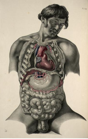
Dissection of the thorax and abdomen, shown in situ. Joseph Maclise, 1856.

Dissection of the thorax and abdomen, shown in situ. Joseph Maclise, 1856.

One aspect that I have always brought up is that heart muscle cells essentially don’t reproduce in adulthood. This is why a myocardial infarction leaves a permanent fibrous scar, that will remain for life. The heart cannot heal back to its normal functionality. And the low proliferative capacity of the heart muscle cells is probably closely linked to the strange phenomenon that they never give rise to cancer.

Alas, this has changed. A paper appeared just a few days ago from the Frisén laboratory showing that heart muscle cells do proliferate, but slowly. About half of them are replaced at some point during adulthood. The technique used to demonstrate this is based on the detection of carbon-14, levels of which increased dramatically in the entire atmosphere of the earth in the 1950’s and 60’s due to test detonations of nuclear bombs. The DNA in heart muscle cells from people who were already adults at that time was found to contain far more carbon-14 than expected, indicating that new cells had been formed.

Science has a podcast with Jonas Frisén where he explains what it’s all about.

When I was in high school, I used to wonder what it felt like to be a teacher, when old knowledge was proven false. Suddenly, I thought, a lot of past work would seem counterproductive.

But this is not how I feel at all! It’s a delightful sensation, a feeling of moving forward.

This reevaluation is a reminder that all our knowledge is provisory, and can be overturned at any time by new evidence. I shudder to think of a world where we didn’t question our old beliefs.


Reversible apoptosis: cancers can postpone the point of no return

April 21, 2009

ResearchBlogging.orgApoptosis – programmed cell death – is one of those processes that are only supposed to go one way. Once begun, there is no turning back. The cell begins to degrade itself through the activation of caspases, proteolytic enzymes that cleave the structural elements of the cell, leading to a series of morphological and biochemical changes. The cell digests itself from the inside, turning itself to a soup of dead organic molecules inside the cell membrane. In the end, it fragments neatly into membrane-enclosed bags that are small enough for nearby cells to phagocytose, i.e. eat up.

Evidence for reversible apoptosis
In a recent paper in the British Journal of Cancer, Tang et al. show that apoptosis may be reversible. They have taken a panel of several cancer cell lines and treated them with an inducer of apoptosis for a few hours, and then washed it off. When they monitored the cells over the next hours, they found that many cells transiently exhibit morphological signs of apoptosis: they shrunk, the nuclei condensed, and mitochondria were degraded, but a few hours later the cells returned to their normal shape. The limiting event from which the cells had no chance of return was when degradation of the nucleus had begun. Caspases were activated at the same modest level from 3 up to 48 h after induction, even though the cells were back to their normal shape before 24 h.

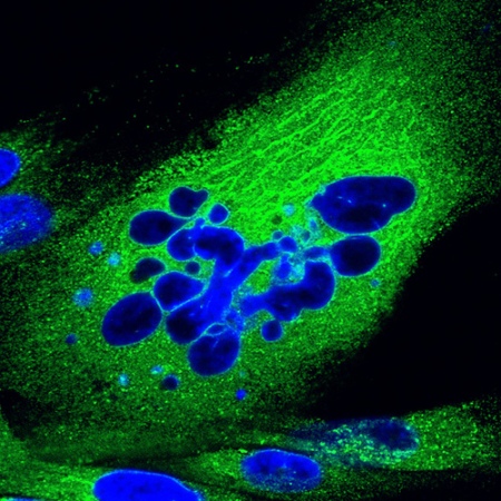
Human fibroblast undergoing nuclear fragmentation in a late stage of apoptosis. The nucleus is stained in blue and the cytoplasm in green. Photomicrograph by Joerg Schroeer.

Human fibroblast undergoing nuclear fragmentation in a late stage of apoptosis. The nucleus is stained in blue and the cytoplasm in green. Photomicrograph by Joerg Schroeer.

Loss of bistability in the apoptosis signalling network
In terms of the signalling network, apoptosis should be a bistable process. The cell should very firmly be either apoptotic or non-apoptotic. Cells that start to digest themselves and then stop should have a strong fitness penalty. We would expect the evolutionary process to select strongly against a reversibility trait both in an organism and in a group of cells. Contrary to this expectation, the reversibility trait predominates in all the cell lines investigated in this study. Something has been lost, or gained, in their signalling network. But how can they survive a partial self-degradation?

Mitochondria – redundant in cancer cells?
The hallmarks of apoptosis that are investigated in the present study reflect degradation in three steps: First, the cytoskeleton, leading to cell shrinkage. Then, the mitochondria, shutting off the cell’s aerobic metabolism. And finally, the nucleus, with the entire gene regulatory system. But the cytoskeleton is made to be remodelled, so it’s not that surprising that the cells can cope. With the mitochondria, it’s a different story. Cancer cells almost universally switch to anaerobic metabolism during progression. This is known as the Warburg effect. It is a very curious phenomenon that still awaits explanation. Many of the mitochondria in tumour cells therefore lack any useful function. In a sense they are little more than decoration, present mainly for historical reasons.

The hen and the egg
Is it the case that tumour cells first acquire apoptosis resistance through mutations that repeal the bistability of their signalling network, and then are forced to rely less on their mitochondria because they are sometimes degraded? Or is it the other way around? Or are these two processes driven by unrelated factors, leading to reversibility of apoptosis by chance, as it were?

It is too early to draw any conclusions. Further studies should investigate reversibility in relation to biochemical and not just morphological parameters. I will write more on the properties of bistable signalling networks and their robustness during the coming week.

Full reference:
Tang, H., Yuen, K., Tang, H., & Fung, M. (2008). Reversibility of apoptosis in cancer cells British Journal of Cancer, 100 (1), 118-122 DOI: 10.1038/sj.bjc.6604802


A prosthetic motivational system

April 13, 2009

Changing patients’ behaviours is one of the most challenging parts of medical practice. It is hard to overstate how much of our disease burden would simply disappear if people would not smoke, drink alcohol in moderation or not at all, exercise regularly and eat healthy food. Perhaps a third of current cancer morbidity would cease to be, and far more of the cardiovascular morbidity. Of course, people will die from something in the end, but the gain in terms of productive life years would be enormous.

The reason why it’s so difficult to change people’s behaviour is that the reward systems of our brains are such powerful regulators of what we do, and it’s frustratingly difficult for any physician to override the patient’s urge to have his next cigarrette.

When there’s an important problem in the world, there’s also a bunch of scientists trying to solve it. Taking control of our motivational systems could transform our societies in profound ways. Can it be done?

Yes, says Christopher Harris. He is an old friend of mine and a neuroscientist at the university of Sussex. He is personally leading a campaign for the use of deep brain implants to control our reward systems – iPlants, in his coinage.

Deep brain stimulation is a technique similar to the pacemaker. Electrodes are placed in a specific brain region, which can then be activated by passing current through the electrode.

The neuroanatomy of reward is very well known. A small group of nerve cells in the midbrain, when stimulated, release dopamine throughout the entire prefrontal cortex, which is our decision generator. Deep brain stimulation to control reward would be very similar to its application against Parkinson’s disease, in which dopamine signalling is impaired, leading to symptoms of the motor system. Thus, the technology is tried and tested in humans.

The human motivational system has been shaped over millions of years of evolution to a degree of robustness, which is why we find it so difficult to change. Sweet food is an instant reward for most people, as are alcohol and many drugs. The modern society has developed spectacular shortcuts to dopamine release, with the unfortunate effect of making many people’s lives less functional. Obesity and addiction are long-term scourges caused by the inability to resist short-term dopamine stimulation. Here is a technology that could change all that.

But who will push the button?

Improperly used, a system like this might make the patient a slave to the man with the remote control. And supposing that an accountable system can be put in place to prevent that, there is the problem of how, exactly, to connect stimulation to the desirable behaviour. Christopher suggests:

Physical exercise can be motivated by repeatedly delivering rewarding brain stimulation (RBS) whenever an animal runs on a treadmill[10] or lifts a weight[11]. iPlant-driven exercise programs would apply the same principle to humans, for example by delivering RBS whenever the user pulls a stroke on a rowing machine or when pressure-sensitive shoe hit the ground during running (see top image). Every exercise program must have a strict time-limit agreed on in collaboration with a physician.

The science and technology of the iPlant is described in some detail on Christopher’s own web page. Whether it will be tried or not is an open question, but it’s not science fiction. All the technology is there, and it works in laboratory rats.

Whether or not the iPlant will be tried, it is clear that our increasing understanding of the brain is driving remarkable advances in technology to modulate and control its function. If we manage these developments properly, we will be able to solve very many problems. But the iPlant also highlights that these advances will bring ethical issues with them of which the general public needs to be aware.


Christopher has put up a few video clips on this topic, more can be found on youtube or on his website.


Remarkable fitness gain from self-organising properties of cancer cells?

April 8, 2009

ResearchBlogging.orgNeuroscientists have their Aplysia, geneticists have their Drosophila. We, in the field of cancer research, have HeLa – the cervical cancer cells of Henrietta Lacks, probably the most widely used cell line in the world. HeLa, and thousands of cell lines like it, form the bulk of our experimental material. Cell lines are made up of cells that have become immortalised through tumour progression, and can be cultured and passaged indefinitely. Most of them grow nicely on glass or on the coated insides of plastic culture bottles, forming a flat monolayer.

Very much of our knowledge about cancer comes from these model systems. But how well do they resemble an actual tumour?

There is a growing recognition that a tumour is a complex organ made up not only by the tumour cells themselves, but also by many kinds of supporting cells – stromal fibroblasts, blood vessel cells, etc – that enable the cancer cells to grow and proliferate. The 3D interactions between tumour cells and the extracellular matrix (that is, all the stuff that makes up tissues but lies outside of cells) are immensely important in determining the growth patterns of the cancer cells.

The tumour organ, if you wish, is possible only because of the strong self-organising properties of the diverse tissue components. Supporting cells line up next to the cancer cells. Blood vessels grow into areas that are poorly oxygenated thanks to signals that are basically the same as in healthy tissues, for example when muscles grow and need a greater blood flow.

It could be said that the tumours have subverted these normal processes for their own malignant purposes. Or, alternatively, you might argue that it is the normal tissue that drives tumour progression, leaving the cancer cell relatively innocent. (These two viewpoints appear contradictory at first, but a moment’s reflection reveals that it only seems this way because the concepts of purpose and guilt have been introduced in spite of being meaningless in this context.)

A recent paper by J Daubriac et al, in Cell Death and Differentiation, investigates a particular kind of self-organisation: when cancer cells that float freely attach to each other and start to organise into a small tissue.

A very special habitat for cancer cells, which particularly concerns mesothelioma researchers like myself, is in the fluid of the thoracic and abdominal cavities. As you might expect, there is always a small amount of fluid surrounding the lungs and the intestines, to reduce friction when they move. The surfaces are made up of a thin layer of mesothelium, which has a lubricating side facing outwards toward the opposite surface.

Occasionally, mesothelial cells come of the surface and start to live as free-floating cells in the fluid. It is thought that they can spend some time there and then settle again if they find a free spot, thereby contributing to healing any wounds in the mesothelium.

Life as a “floater” is quite different from life on the surface, because on the surface it is necessary for the cells to have polarity. Polarity is the essence of epitheliality, and means that there is a distinct surface facing downwards and to the sides (basolateral), and an apical surface with completely different characteristics facing towards the lumen, or hollow space, of whatever the epithelium in question is lining, be it an airway, a milk duct, or an intestine. The basolateral side is characterised by specialised adhesion structures; desmosomes and adherens junctions to other cells, and integrins and cadherins to the underlying connective tissue, usually a specialised basal lamina. The apical side is where secretion and absorption take place, and where specialised structures like cilia and microvilli are found.

Polarity is lost when the cell floats alone.

The cell must have contact with other cells in order to establish the adhesion structures that define its polarity. Normal “floaters” never achieve this. The presence in the thoracic fluid of a group of cells that adhere to each other is an ominous sign of cancer. Most cells, in fact, are programmed to commit suicide if they lose attachment, in a particular type of apoptosis termed anoikis. Cell attachment is a bit like the safety handle on a garden shredder; the moment you stop pressing it, the machine shuts off.

Dr Daubriac has investigated this behaviour by cultivating mesothelioma cell lines in flasks where they could not attach to the bottom. Instead, they started attaching to each other. They quickly reestablished their polarity, forming spheroids in the µm size range. Some of the cells even constructed a basal lamina in the centre of the spheroid, thus recapitulating the normal tissue features rather fully.

Two particularly interesting findings appeared when the spheroids were compared to normal monolayer culture. Firstly, their growth rate was lower. Cells in spheroids appeared to become rather content with sitting there, with neighbours on all sides, and stopped proliferating. Secondly, they were also much less likely to undergo anoikis than monolayer cells. Daubriac and his colleagues investigated the intracellular signalling pathways in some detail and were able to say that those signals that normally lead to anoikis were shut down in spheroid-growing cells.

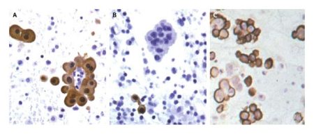
Cytological specimens of mesothelioma cells growing in a spheroid pattern, termed papillary structures. The image is from www.histopathology-india.net/MesoCyto.htm.

Cytological specimens of mesothelioma cells growing in a spheroid pattern, termed papillary structures. The image is from http://www.histopathology-india.net/MesoCyto.htm.

So what is it with these tumour cells that makes them think that they have to build a new tissue? Have they acquired genetic changes that turn this program on under inappropriate conditions?

The authors’ idea is that spheroid growth is motivated by the protection against anoikis that it brings. This implies that there is a selection effect. The cells that could not form spheroids may have died and disappeared much faster, leaving the spheroids to make up a growing proportion of the floating cells. In this case, the decreased death rate may offset the decreased proliferation rate of spheroid-growing cells, leading to a net fitness gain for these cells. Furthermore, the spheroids were able to start proliferating again when they were returned to a normal culture flask and could grow out as a monolayer. It brings an image to one’s mind where spheroid cell groups are the agents of metastasis. If that is so, then a treatment directed against the spheroids might be highly valuable!

Again, we can witness the enormous explanatory potential of the evolutionary paradigm. And it always helps to keep in mind that tumours are among the most dynamic evolutionary systems that exist, because of their widespread genetic instability.

If self-organisation in spheroid growth contributes to metastasis, then it’s a bit revolutionary, because self-organisation usually works the other way! The more the cancer cells try to build a functioning tissue, the less malignant will the tumour be. Cancer cells resembling the original tissue are called well-differentiated, and then there is a whole scale with anaplastic at the bottom, which means that the cancer cells look pretty much like stem cells with no particular tissue allegiance. The differentiation state is closely liked to the patient’s prognosis.

This paper leads to a clear hypothesis that mesothelioma cells have an evolutionary fitness gain from spheroid growth. In principle, that should not be too difficult to prove or refute. I look forward to more research on the topic!

Full reference:

Daubriac, J., Fleury-Feith, J., Kheuang, L., Galipon, J., Saint-Albin, A., Renier, A., Giovannini, M., Galateau-Sallé, F., & Jaurand, M. (2009). Malignant pleural mesothelioma cells resist anoikis as quiescent pluricellular aggregates Cell Death and Differentiation DOI: 10.1038/cdd.2009.32

A single-cell organism that communicates using light signals

April 7, 2009

Some multicellular organisms emit light in a conspicuous way. Fireflies carry beacons that shine in the night, and some deep-sea fishes use phosphorescent appendages to attract prey. However, the information content in these light emissions is probably no greater than “I am here”, or possibly “I am moving in a certain direction with a certain velocity”.

A few days ago, a paper appeared in PLoS One showing that the unicellular organism paramecium caudatus uses light signals of a specific wavelength to communicate. These signals influence the proliferation rate of the protozoa and imply that they not only have a sense of vision, but also a signal-generating organ, and an apparatus to translate the visual input into a representation of its environment, which in turn guides the organism’s behaviour.

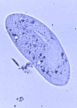
Paramecium caudatum. Image borrowed from A Blog Around the Clock.

Paramecium caudatum. Image borrowed from A Blog Around the Clock.

I found this story through the most interesting Neurotypical blog, written by a neuroscientist who starts off almost apologetically by saying that this has nothing to do with neuroscience. I beg to differ.

Although the paramecium possesses no nervous system, it clearly has all the necessary faculties for information processing of the kind with which neuroscience concerns itself. It is not the substrate that determines the dynamics of an information processing system, but the structure and organisation of the network.

Check out the post by Neurotypical, as well as the the original paper, it’s open access!


Carnival of Evolution

April 2, 2009

coebutton

This months carnival of evolution has just been published at the Oyster’s Garter. It’s an unusually nice compilation, which I can freely say since I’m contributing this time.

Check it out!


Lungs – what are they really good for? More on lungless amphibians

March 27, 2009

ResearchBlogging.org This is a follow-up on yesterday’s post, which discussed a lungless frog species recently discovered on Borneo. Victor H. Hutchison has written a comment in the journal Current Biology that highlights a few interesting concepts.

Sometimes I give an introductory lecture on the histology of the human lung to undergraduates in our department. I invariably start off by asking the students what the organ is good for, and I always get the same two answers: Gas exchange and barrier function. I never realised that there is a third function in many animals, although it’s obvious when you think of it: flotation. As a submarine can regulate its buoyancy by filling tanks with either water or air, so can many amphibians regulate theirs with their lungs.

Hutchison explains that amphibians may be particularly susceptible to losing their lungs because they have rather inefficient breathing dynamics. Apparently, they cannot breathe by changing the volume of the thorax with muscles, like we do. Instead, thay have to force air into the lungs by a swallowing motion. Then, because of the higher pressure built up in the lungs, expiration takes place when they open their mouths again. Further factors that contribute to the redundacy of lungs are a higher body surface area to volume ratio, a permeable skin with capillaries growing into the epidermis (unlike ours, which stop in the underlying dermis), and low metabolic rates due to cold temperatures.

A unifying trait for the previously known lungless salamanders and the recently discovered lungless frog is that they live in cold streams. Hutchison therefore proposes that the loss of lungs helps keep these animals on the bottom, preventing them from being swept away by the water. He does not mention that gas exchange will be much more efficient in moving water than in a still-standing pond, but this seems an obvious observation to me.

Some amphibians have reduced lungs while still retaining the capacity to use them. One example is the Titicaca frog (Telmatobius culeus). This animal lives in high-altitude waters in the Andean mountains. Normally, these frogs stay underwater and use their many skin folds for gas echange. These folds have very superficial blood vessels and are ventilated by a “bobbing” motion. In addition, the frog’s blood is very rich in hemoglobin.

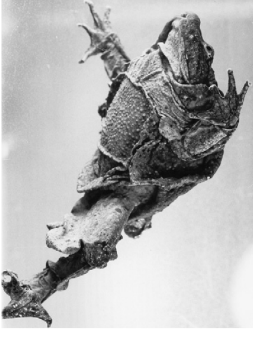
Titicaca frog. Image from Hutchison's paper.

Titicaca frog. Image from Hutchison's paper.

Hutchison remarks that there are probably more lungless frog species with specimens sitting around in museums of natural history, that have never been dissected. Perhaps these museums would be helped by a small CT scanner? (More likely perhaps, they would be helped by people with an interest in going through their vast collections and cataloguing them.)

Full reference:
HUTCHISON, V. (2008). Amphibians: Lungs’ Lift Lost Current Biology, 18 (9) DOI: 10.1016/j.cub.2008.03.006


Breathing in the bitter cold: lungless frogs and a fish without erythrocytes

March 26, 2009

ResearchBlogging.orgToday I stumbled upon two blog posts that really capture some of the beauty of the diverse adaptations in nature.

Random Biology writes about creatures living in cold waters. Water can carry oxygen at a far greater density at lower temperatures. This simple phenomenon, combined with the slower metabolism of cold tissues, has made it possible for certain salamanders to get along fine without lungs. All their breathing occurs through the skin.

In a recent paper in Current Biology, David Bickford and two colleagues describe the same phenomenon in a frog! It’s called Barbourula kalimantanensis, and lives in Borneo. Interestingly, it has apparently retained the lining of the lungs and thoracic cavity, called the mesothelium.

Barbourula kalimantanensis. Image from the paper by Bickford et al.

Barbourula kalimantanensis. Image from the paper by Bickford et al.

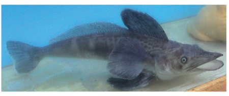
In a still more fascinating post, Biochemical Soul describes a fish with a new way of dealing with freezing temperatures. Or several, actually. It is the Channichthyidae family of icefishes, which live in Antarctic waters that are often below freezing point (but still liquid, of course, because of their salinity). These fishes have no hemoglogin, and consequently no red blood cells. They also lack myoglobin, the related molecule that stores oxygen in muscle cells. They rely instead on the greater oxygen-carrying capacity of their cold blood. With no erythrocytes, the viscosity of the blood decreases, which helps circulation. And to compensate for the lack of oxygen carriers in the blood, they have a 4-5 times increased stroke volume of the heart. This was originally described in 2006 by Thomas J. Near and coworkers in an open-access paper in Molecular Biology and Evolution.

Icefish. Image from the paper by Near et al.

Icefish. Image from the paper by Near et al, referenced below.

Cool stuff! (Don’t excuse the pun.)

Full references:
BICKFORD, D., ISKANDAR, D., & BARLIAN, A. (2008). A lungless frog discovered on Borneo Current Biology, 18 (9) DOI: 10.1016/j.cub.2008.03.010
Near, T. (2006). A Genomic Fossil Reveals Key Steps in Hemoglobin Loss by the Antarctic Icefishes Molecular Biology and Evolution, 23 (11), 2008-2016 DOI: 10.1093/molbev/msl071


Is the Central Limit Theorem an engine for biological stability?

March 25, 2009

Biological information systems, like any others, struggle constantly with randomness. Our bodies are precision instruments to measure very many things at the same time – light, vibrations, gas pressures, concentrations of salts and hormones, to mention a few. Any of these measurements can be thought of as a sample. Now, randomness can cause the sample to lie quite far off from the actual measure. A possible solution is to resample the sample! This is not intuitive, and I will explain it below. Perhaps this is the reason why many signalling pathways in biology have so many links in the chain from receiver to effector!

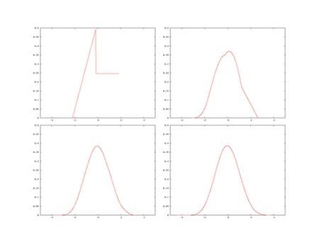
The Central Limit Theorem states that if you draw a sample from a population and calculate the mean of the sample, and then repeat it several times, the means will form a normal distribution around the true mean of the original population. This means that even if the original population has a wild distribution, repeated samples of the population come closer and closer to the true mean.

Take a look at this example to see what it means:

Image from Wikipedia

Image from Wikipedia

Here, the original distribution is on the top left – highly irregular. But if we take samples of two numbers at a time from this distribution and plot their means, we end up with the distribution on the top right – already a great step towards normality! With three and four in the sample, we get the bottom left and bottom right, respectively.

Nearly all cell surface receptors signal through a pathway of messenger molecules. Not just one, but a whole cascade. The traditional explanation for this phenomenon is that the signal can be easily amplified in this way. But perhaps the real driver is the stability of the readout that can be gained. There are similar organisational features in other places too, for example in the transmission of visual information from the retina. The signals pass through a few serially arranged neurons on their way to the visual cortex. Perhaps this is what prevents our field of view from flickering? (The rods are exquititely sensitive and can detect a single photon.)

Perhaps I should write this up and submit it to the journal of Medical Hypotheses? (This is one of the few scientific journals that require no proof whatsoever, and as a result the journal contains everything from well-supported testable hypotheses to completely far-out ideas, such as the benefits of masturbation against nasal congestion.)

What do you think? 🙂


Surely you’re joking, mr Ernberg!

March 20, 2009

Have we really solved the riddle of cancer? Yes, says Ingemar Ernberg, the venerable professor who has written the foreword to ”Prostatacancer”, hot off the presses of the Karolinska University Press.

I was somewhat surprised by his argument, which runs something like this: If there ever were a riddle of cancer, we have solved it by showing how the cell’s actions are controlled by gene regulatory networks. With ceaseless environmental perturbations of these networks, coupled with the powerful organizing principle of evolution, nothing mysterious remains.

Certainly, the advances in tumor biology have been tremendous over the past decades. And it is no coincidence that much of what we have learned about genetics and cell signaling has been discovered in the context of cancer. But can we really say that we understand these processes because we have identified the constituent parts and some of their connections?

If this were true, the riddle of consciousness was really solved in the 1800:s, when Golgi invented the silver staining that for the first time enabled us to see how neurons connect with axons and dendrites.

What professor Ernberg does not consider is the complexity that arises through the dynamic information transfer of the network. On this higher-order level, in cells just as in the nervous system, behavior emerges that cannot meaningfully be accounted for by cataloging the interactions of the component parts.

If this is not immediately obvious, consider the following. Certain genes, when upregulated, cause cells to proliferate a lot. An example is the c-myc gene. This gene can be accidentally moved to the place for the immunoglobulin gene in certain lymphocytes when they are infected with the Epstein-Barr virus. As a result, the lymphocytes proliferate enormously, and we have leukemia. Other genes, which sometimes cause cells to proliferate a lot, can also sometimes cause them to die a lot. An example is th JNK gene. There has been much controversy over whether JNK is pro- or antiproliferative. Now, it is generally accepted that it is both.

In total, we humans have around 20 000 genes. Even if each gene only interacts with 10% of the other genes, and the interaction is always linear, a model to explain the cell’s behaviour would be totally intractable even with enormous computing power. When many of the interactions are non-linear, it becomes clear that a successful description of this system, with the power to predict what it will do, must consider a higher level of organisation. Analogies abound; reading the Pickwick papers by Charles Dickens letter by letter vs. by the meaning of phrases and their conjunctions (D. Hofstadter, in Gödel, Escher, Bach), or understanding a city by copying the telephone directory vs. actually finding out where people are going every day and why (Sidney Brenner).

The riddle of cancer remains. The most important discovery we have made so far is that the riddle of cancer is identical the to riddle of Life itself; namely how the genes and proteins that are the basic units of biological information, as well as the basic operators on this information, together determine the fate of the cells which are the smallest units of life as we know it.

(I am indebted to professor Ernberg for having created much of the intellectual arena where I have encountered several of the more groundbreaking recent advances of thought in tumor biology, and I argue against him safe in the knowledge that he will only be pleased that his ideas are debated.)