Will your cell phone give you CANCER?

March 17, 2009

The world is full of hidden dangers, in some people’s view. Some things that were thought harmless at some point in history have turned out to be genuine killers – lead, tobacco smoke, and asbestos, to mention only a few examples.

Our historical success in identifying unobvious dangers has lead some people to develop mental models where there is always a widespread behaviour or exposure that is next in line to be uncovered as a cause for illness and mortality. Others believe (as I do) that with time, the remaining environmental hazards to be discovered will be less and less important, because the size of the adverse effect correlates strongly to how easy it is to detect, and therefore most of the really dangerous stuff is probably already known.

Whichever attitude people take, most agree that it is best to base specific recommendations on a dispassionate reading of the scientific data. However, opinions differ greatly about how strong the evidence has to be in order to restrict something that might be dangerous. “The principle of caution” states that it’s better to regulate when we are uncertain. But how uncertain?

A useful guide for determining the strength of evidence has been proposed by Austin Bradford Hill (1965). Its criteria have been summarised as follows: Causality becomes knowable when scientific experiments demonstrate, in a strong, consistent (repeatable), specific, dose-dependent, coherent, temporal and predictive manner that a change in a stimulus determines an asymmetric, directional change in the effect.

In this post, which I fear will be rather lengthy, I will try to review the evidence for and against dangers of mobile telephones. The positive health effects are significant (easier access to medical advice and emergency services, in particular), but we will only look at the possible risks here.

800px-several_mobile_phones

There is a debate over the health risks of electromagnetic radiation in general. When we use a mobile telephone, we expose ourselves to an electromagnetic field which penetrates about 4 cm into the head. Some national authorities have defined exposure limits. For example, the Swedish Radiation Safety Authority requires that all phones cause less energy absorption than 2 W/kg in human tissues, a limit that has been more or less arbitrarily chosen on the base of acute radiation effects. Absorption of radiation causes tissues to become warmer, and 2 W/kg is far below any measurable thermic effect. (This is the general principle of the microwave oven, and the only completely indisputable biological effect of electromagnetic fields.)

Other dangers have been seriously investigated, particularly the risk of cancer and of neurological disorders such as headaches, dizziness, and dementia.

Loud activist groups are lobbying for more restrictions. The most authoritative critics are probably the BioInitiative Group. They released a report in 2007 urging the prohibition of strong electromagnetic fields. I purports to “document serious scientific concerns about current limits regulating how much EMF is allowable”. But although scientists are among the authors, the document is not a piece of scientific criticism. It is a jumble of cherry-picked research reports and unfounded claims. The authors are quite consistent in only mentioning research that supports their own point of view. Furthermore, they accept controversial data regardless of prior probability.

Let’s say that I were to investigate whether my zodiac sign is a strong risk factor for Parkinson’s disease. If I were to choose a confidence level of 95%, I would end up being wrong 5% of the time, which is the usual cutoff in science. Now, let’s say I get a positive result. Does that mean that sagittarians are at increased risk for Parkinson’s? No, because I have to take into consideration the prior probability that my result is correct. And our current understanding of disease physiology and the differences between people born at different times in the year makes such a result utterly implausible, at best. This is, incidentally, why it is impossible to prove that homeopathy works without first changing our understanding of the laws of chemistry.

The BioInitiative report, fearless of ridicule, is thus forced to conclude that since adverse effects of very low intensity electromagnetic radiation have been seen in cells in certain experiments, it must be the information conveyed by the radiation rather than the heat in the tissue that causes damage. It seems to be envisioning “death rays” that can be specifically tailored to destroy tissues, and that coincidentally are similar to the electromagnetic fields that surround us every day.

No, the BioInitiative report is not serious. What, then, do the big public agencies have to say?

The WHO has concluded that there is no increased risk for cancer or any other diseases, but that cell phones may lead to traffic accidents and may interfere with pacemakers. The EU:s expert group has concluded that there is no increased risk for cancer except possibly for a benign tumour called acoustic neuroma, and only for these who have used the cell phone for more than ten years, with almost a doubling of the incidence rate.

Here is where risk communication comes into the picture. About 80-100 cases of acoustic neuroma are diagnosed every year in Sweden, with a population around 9 million people. The risk is consequently about 1/100 000 for a person in a year. An increase in the relative risk by 100% corresponds, then, to an increase in the absolute risk from 0.001% to 0.002%.
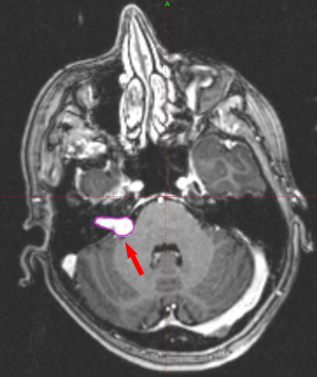
MRI image showing an acoustic neuroma

MRI image showing an acoustic neuroma

And this is the most tangible risk. With regards to other types of cancer and neurological diseases, risks completely fail to pass the Hill criteria. The effects, if any, are weak, inconsistent, unspecific, weakly dose-dependent if at all, incoherent and unpredictive. Sadly, space does not permit me to go through the entire literature in this post, but good summaries are available from the WHO and the EU.

In spite of the lack of evidence, the campaigners are making headway. In September 2008, MEPs voted 522 to 16 to urge ministers across Europe to bring in stricter radiation limits and said: “The limits on exposure to electromagnetic fields (EMFs) which have been set for the general public are obsolete. The European Parliament is greatly concerned at the Bio-Initiative international report which points in its conclusions to the health risks posed by emissions from devices such as mobile telephones, UMTS, WiFi, WiMax and Bluetooth, and also DECT landline telephones. The plenary therefore calls on the Council to […] set stricter exposure limits.”

The politicians have to listen to the people, after all, even when the people are wrong. The whole conundrum is hilariously exposed in this episode of “Yes Minister”. (Go to YouTube for the remaining parts of the episode.)

In my opinion, the moral of the story is not that our elected leaders must stand up for science (although that is also true), but that we must all aid and assist our decisionmakers to behave rationally. If they think people want homeopathy and prohibition of GMO-crops, then that is what we will end up with.

When we have superior knowledge, the democratic right to share it is also a duty.


Self-medication in a caterpillar

March 11, 2009

ResearchBlogging.orgSelf-medication among animals is sufficiently well studied to have a term of its own: zoopharmacognosy. Until now, such behaviour has mainly been observed among higher primates. Chimpanzees with intestinal parasites may seek out and eat shoots that are not part of their normal diet – indeed, that are toxic and make them look like they had just bitten deep in a slice of lemon. Sometimes, the same plants are used by local humans in traditional medicine.

Cool, right? But hardly rigorous. What we would really like to see in order to have good proof for the concept is an example of an animal eating a foodstuff whose medical properties can be proven, and furthermore only eating it in times of illness, and preferably suffering negative effects if they eat it when they are healthy.

A woolly bear caterpillar. Image from Wikipedia. NB: probably not the exact same species as in the study in question

A woolly bear caterpillar. Image from Wikipedia. NB: probably not the exact same species as in the study in question

Such a study has been published for the first time today, with a very surprising animal species as the protagonist: the woolly bear caterpillar. This is the larval stage of a moth of the Arctiidae family, which is apparently well known for its habits of communicating by ultrasonic sound waves, generated from a specialised organ.

Michael Singer and his co-authors have infected woolly bear caterpillars with an intestinal parasite and supplemented their diet with plants that contain substances called pyrrolizidine alkaloids (PA:s), but that are very nutrient-poor.

They found that infected larvae fared better if they ate the PA-containing plant-stuff, whereas uninfected larvae fared worse. Fewer parasites survived to reach maturity if they had access to PA. As a group, the caterpillars did not eat significantly more of the PA-food when they were infected, but there was a suggestive trend where the larvae with a greater infection load ate more PA.

In this case, the behaviour is clearly not socially learned, as it may be in the apes, but somehow coded into the neurological hardware of the caterpillar. Or rather, a subset of the caterpillars. If the parasites ceased to exist, the subpopulation most likely to eat PA-containing plants would be at a disadvantage since PA is toxic. Here is another instance of evolutionary dynamics creating a phenotype that is only advantageous in the face of an external threat – just like sickle-cell anemia in humans, which confers a measure of protection against malaria, and many other genetic diseases.

A 2005 Nature paper by the same authors showed that the peripheral taste nerve cells actually change their signalling in infected caterpillars and fire off more signals in response to PA. They speculate that the immunological response to infection is the direct cause of this change, which doesn’t even need to involve the neural cluster that resembles the caterpillar’s brain. From the perspective of information processing, this paper is a small triumph for those who try to find complex behaviours in relatively simple biological organisms.

Singer, M., Mace, K., & Bernays, E. (2009). Self-Medication as Adaptive Plasticity: Increased Ingestion of Plant Toxins by Parasitized Caterpillars PLoS ONE, 4 (3) DOI: 10.1371/journal.pone.0004796

UPDATE:
BOth Carl Zimmer at The Loom and Ed Yong at Not Exactly Rocket Science have written excellent posts on the same research.


The Meaning of Systems Biology

March 9, 2009

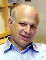
Today, I had the pleasure of meeting prof. Marc Kirschner. He is the chairman of the department for Systems Biology at the Harvard Medical School. It’s a visionary place with about 200 scientists, small by American standards but far larger than my current department.

Marc Kirschner

Marc Kirschner

Kirschner is one of the world’s foremost cell biologists, and one of the first to thoroughly explore the information-processing capabilities of the cell. In his 1997 book Cells, Embryos and Evolution (written together with J.C. Gerhart), he discusses, for example, the capability of the cell to be used as a computing machine, in principle.

I read up a bit before meeting him, and came across an article he wrote for Cell in 2005 titled “The Meaning of Systems Biology”. Here he endeavours to explain what this new field is about, really. And he is the right person to do it, since he is more or less one of the founding fathers of the field.

Rarely have I seen such an established scientist move with such caution in his own field. He describes systems biology as a scientific branch in the making, and argues that only in retrospect will we know exactly what has come of this fruitful entanglement of genetics, molecular biology, cell biology, physiology, and evolutionary theory, all coupled with new high-throughput techniques.

Systems biology is not all about an increased data-generating capacity. It is, in Kirschner’s opinion, also about “a smaller scale view, totally compatible with and partially dependent on the global analysis of high-throughput biology. This view spans in vitro biochemistry to what is now called synthetic biology and it has as its goal the reconstruction and description of partial but complex systems.”

In the end, Kirschner only very reluctantly offers a rather long definition, which he says must remain tentative. At its core, I think it means that systems biology is the study of biological complexity through modelling of the underlying mechanisms, quantitative measurement, and theory.

So how did our meeting go? Well, I pitched a project idea that I’d given a lot of thought, and that is in my own opinion extremely elegant, well-conceived and likely to change the way we see the world. One’s ideas become like babies sometimes if you allow them to.

He thought there might be something in it, and suggested I should talk to the person in his department who has got the methods I would require – incidentally, the only person in the world who does. I certainly will. And then we’ll see where this ends!


The beauty of the villus escalator

March 6, 2009

Today I have lectured for three and a half hours on histology. We have gone through slides of all the major organs, scanned into imaging software that enabled us to go to different magnifications in any part of the specimens.

Going through slides for demonstrations like these is almost always an aesthetic treat. This time, the single most beautiful thing that struck me was this:

duodenum1

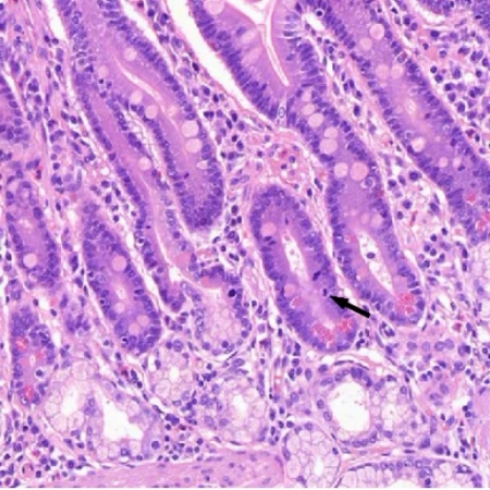
This is a section from a human duodenum, the first part of the small intestine. Lovely, isn’t it?

duodenum21

What we are looking at here is a mitosis at the bottom of a duodenal villus. At the base of the villi, there are progenitor cells, or stem cells if you want, that constantly regenerate and that give rise to all the cells of the villus epithelium. Cells on the apex of a villus will degenerate and die, and will be replaced by new cells moving from the base where they are regenerated.

In essence, the epithelial cells are in a constant but slow motion towards the top of the villus, where they will reach their ultimate demise.

Any cell that thinks differently, and wants to stay, faces a stiff current of upwardly-moving cells that will sweep it along in its never-ending tide.

The epithelial cells are constantly exposed to the slightly toxic cocktail of eaten stuff and microbes in the gut, and even during their short life-time they will be expected to acquire genetic changes. But the constant flux makes it very difficult for any of them to ever develop into a tumour. I wonder if the turnover rate is faster in the small intestine, where tumors are much rarer, and where the energy content of the dying cells is recycled through absorption, compared to the large intestine, where tumours are much more common and only water is absorbed?

The really cool thing, however, is that the villus escalator model has been known for such a long time. Histologists noted more than a hundred years ago that mitoses only occured at the base. This simple observation was sufficient to make the inference that the epithelium is constantly rejuvenated at the base and degenerates at the apex – a model that has been extensivly corroborated since.

UPDATE: This post seems to get lots of traffic from people who look for histological images. Feel free to use the ones in this post for any purpose you like! It’s polite to mention where they came from, though.


Mathematics of a Lady Tasting Tea

March 5, 2009

Here is a gem from the history of science. I happened to come aross this classic 1956 paper, where the mathematician Sir Ronald Fisher explains basic statistical concepts as they apply to a lady who claims she can tell by tasting a cup of tea whether the tea or the milk was added to the cup first.

With hardly any mathematics, but with the stringent logic that is the mathematician’s mark of nobility, he asks and answers the question: how many cups must she identify correctly in order for us to believe her claim?

The answer is eight out of eight, if half the cups are of each kind. Or ten out of twelve. Read it to find out why!

Sir Ronald then proceeds to explain the basics of randomisation in experiments, and of obtaining greater statistical power by increasing the sample size. He pulls it off without using these somewhat technical terms, in a prose whose elegance I admire.

This is some of the best science writing I have seen in a long time.


Why do female doctors have more fun with mammograms?

March 2, 2009

ResearchBlogging.org
Cancer deaths could be reduced by one third, or more, according to expert estimates. One of the strategies to reach this goal is the use of screening programmes. Women in most high-income countries will participate during their lifetime in a screening programme for cervical cancer, and another for breast cancer. Both types of screening have an essential component of image interpretation by a doctor – either a mammogram, i.e. a special x-ray picture of the breast, or a Pap smear, which is a sample of cells from the vagina that is examined under the microscope. These tests are like windows into the magnificent palace of the female body, through which doctors peer like nightwatchmen to try to make out whether any of the shadows in the darkness inside are dangerous lurkers.

Millions of such pictures are examined every year, by doctors suffering under the same unfavourable circumstances as pilots: everything is routine, but the consequences of even a slight error could lead to disaster and death. Decision-making is a challenge. Every doctor must calibrate him- or herself to a set-point where he detects as many as possible of the real positive cases without over diagnosing and creating false positives, which will cause patients to needlessly undergo maiming surgery. Doctors’ decisions, like anybody else’s, are affected by such mundane factors as their alertness, their mood, their serum caffeine levels, and the weather.

This subjectivity needs to be managed.

Of course, doctors have lots of strategies to do that. Most of the time, it works very well. Doctors meet, discuss cases in rounds, and recalibrate their set-points. The keep the images and monitor the patients for decades to see whether any tumours develop that they could have detected earlier. And they do research into the predictors of accurate diagnosis.

One such paper appeared last week in the American Journal of Roentgenology. It investigates the very plausible hypothesis that doctors who enjoy their work also diagnose more accurately. Berta Geller and her colleagues sent a questionnaire to 131 breast radiologists, and then linked their answers to known performance on 700 000 mammograms in a database. In the end, there was no significant connection between the reported enjoyment of interpreting mammograms and the performance of the doctors. This finding is very reassuring and in my opinion a bit surprising.

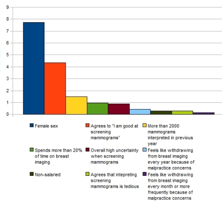
But these is more: The authors have investigated what factors are most likely to predict doctors’ enjoyment of interpreting mammograms, and these are the results:

Predictors of enjoyment

The y axis shows the odds ratio, which can be understood as “fold change in likelihood”. As the diagram shows, doctors were roughly eight times more likely to enjoy interpreting mammograms if they are women! This was a much stronger predictor than the feeling of competence, which comes next. The most important negative predictor was the fear of malpractice suits. (This is a US study.) The “Non-salaried” bar in the middle compares doctors who are paid per case to those paid a fixed salary. Not much difference there.

Why on earth do women find the work so much more enjoyable? There were 101 men and 29 women among the doctors, which should be enough to keep random variation out of question. (That adds up to 130 doctors – one is missing, who perhaps did not state his/her gender.)

Are there hidden confounders? Perhaps the women were younger on average in this sample, and more enthusiastic? No, because older doctors enjoyed their job more than younger doctors did. Could it be that women feel disproportionately skilled at interpreting mammograms? Or is it in fact a real difference, due to some other factor such as that all the doctors identify more with female patients, or that women enjoy image interpretation more in general?

All rather unlikely explanations I think. But science is at its best when it reveals unlikely chains of causation to be true! I hope someone will make the effort to find out whether this is a consistent finding and, in that case, what causes it.

Full reference: B. M. Geller, E. J. A. Bowles, H. Y. Sohng, R. J. Brenner, D. L. Miglioretti, P. A. Carney, J. G. Elmore (2009). Radiologists’ Performance and Their Enjoyment of Interpreting Screening Mammograms American Journal of Roentgenology, 192 (2), 361-369 DOI: 10.2214/AJR.08.1647


Explaining cancer dynamics with game theory

February 28, 2009

 Building a tumour takes teamwork!

Isolated groups of few cancer cells generally do not give rise to new tumours. Micrometastases, i.e. very small clusters of tumour cells, can frequently be found in the lymph nodes and bone marrow of cancer patients. These micrometastases tend to lie dormant after removal of the primary tumour – often for the remainder of the patient’s life. Moreover, it is well known that much cancer surgery leads to a veritable spraying of cancer cells in the operation wound, but it is very rare that these cells form any metastases where they land.

It is only a few years since the mainstream view of cancer researchers turned towards viewing the tumour as a complex society of cells, including tumour cells of heterogeneous natures, supporting stromal cells, and ingrowing blood vessels. These cells all have to work together in order for the tumour to grow and progress. It is a hotly debated issue whether the stromal and vascular cells are deceitfully co-opted by tumour cells which secrete growth factors similar to those occurring e.g. in wound healing, or whether the non-cancer cells surrounding the cancer cells are in themselves supporting and accelerating the tumour growth and progression.

It is in this complex tissue interplay that the tumour cells live and evolve. Cancer cells are genetically dissimilar from the people that are their hosts and progenitors, because they accumulate genetic changes of various kinds – mutations, deletions, amplifications, translocations – that enable them to proliferate independently and so on. One common first step in carcinogenesis is a genetic destabilisation, which leads to a much increased rate of genetic change. Hence, tumour cells constantly develop new genotypes and phenotypes and grow into a heterogeneous society of cells.

Now, in almost every situation where cooperation exists, dynamics come into play that are described by game theory. If everyone cooperates, it is typically advantageous for a single organism or cell to defect and reap the rewards of everyone else’s cooperativity while not contributing. This incentive to defect can lead to the collapse of the cooperative system, or to the emergence of a counter-strategy which rewards cooperators and punishes defectors. Against these strategies, in turn, there are other counter-strategies, and so the play of Nature continues. We can observe it in populations of bacteria, honey bees, wolves, and humans. And in cancer, or so we expect.

But what does it mean for a cancer cell to defect, and cease to cooperate? For about ten years, scattered groups of scientists have been building models where tumour cells of two different kinds derive certain benefits and drawbacks from being next to other cells of the same or of the other kind. (See this paper for a review.) Models like these are great fun, and they highlight the importance of evolutionary dynamics within the tumour. But thus far, the fitness modifiers have been very arbitrarily chosen, and can hardly be said to represent the physical realities of the tumour except on a very abstract level.

There are two conceptual problems that make it very difficult to conceive of what defection in a tumour cell would look like. The first is that the cancer cell bathes in the same soup of growth factors as its immediate neighbours, and many of the stimulatory signals that are emitted by the cancer cell end up targeting the cell itself – so-called autocrine stimulation. This means that if the cancer cell were to stop stimulating its neighbours, it would also stop stimulating itself, and that would hardly ever result in any fitness increase. The second problem is that the cost of signalling is very low, and most of the cooperative behaviour takes the form of signalling through secreted molecules. That means that even if another cell has something to lose if the first cell stops its signalling, the first cell has almost nothing to gain. (Anti-proliferative signalling by tumour cells is very uncommon. I do not know of any proven example.)

In a recent paper, D. Basanta and co-workers suggest a biochemical basis for an act of defection, coupled to game-theoretical interactions. Their model brings in the Warburg effect, which is common to tumours of nearly all kinds. It consists in a shift from aerobic to anaerobic metabolism – great for when the blood supply is strained, as it usually becomes when tumours grow beyond a few millimetres in size. According to their reasoning, cells that shift their metabolism in this way adversely affect their neighbours, because they release toxic metabolites. They proceed to construct a set of conditions under which invasive tumour cells start to migrate out from the tumour after sufficiently many cells have turned anaerobic. In fact, they provide a completely new theoretical framework that may explain in evolutionary terms why the Warburg effect is so ubiquitous.

As good theory should, this model yields predictions that are testable in principle. Unfortunately, it is not really possible to eliminate locally the toxic by-products of anaerobic metabolism, or to prevent the Warburg effect from appearing at all. While we wait for experimental science to catch up, these theoretical models continue to help us form a tentative understanding of the principles behind the tissue interactions in tumours.

Update: The Carnival of Evolution has just been published in its ninth edition at Moneduloides, and this post has been included!


Nature endorses science blogging

February 27, 2009

The latest issue of Nature carries an editorial encouraging scientists to blog about their research. It discusses how to relate to public discussion of unpublished results, and ends by saying:

”[…] there are societal debates that have much to gain from the uncensored voices of researchers. A good blogging website consumes much of the spare time of the one or several fully committed scientists that write and moderate it. But it can make a difference to the quality and integrity of public discussion.”

This is obviously something to keep in the desk drawer for any scientist who keeps a blog and who may run into a discussion with colleagues or department heads about whether it is valuable to spend time on writing about science in this format.

However, good science blogs have been around for years. The reason why this endorsement comes now is probably the joint impact of the blogosphere and preprint servers. In mathematics and physics, it is very common to upload manuscripts to arxiv.org before they are submitted to peer-reviewed journals. Nature has started a similar preprint server for the biological and medical sciences. These preprints may be discussed by the authors on their own blogs, or by other readers in the scientific community. In the past, the same kinds of discussions would occur only in the physically limited space of conferences or through personal contacts. Now, discussions about new science can be carried out before the eyes of the world, with links directly to the findings so that everyone can make their own interpretation. The monopoly of the scientific journals is evaporating. Nature appears to have realised now that this is a development they cannot hinder, and therefore they reluctantly accept it. Seen from this perspective, the editorial represents a walk-over victory for open science!


I can see through your forehead!

February 24, 2009

Well, what to make of this? The Zooillogix blog alerts me to a new report on the barreleye, a bizarre deep-sea fish. It has recessed its eyes quite deep underneath the skin of the forehead, which is completely translucent! Over the mouth are two small dots that look like eyes, but are in fact nostrils of a sort.

Check out this video:

Deep sea news has more coverage.


How cells decide to live or die: an ambitious effort at the MIT to map the wiring

February 24, 2009

At the first glance, it may not be obvious why a cell should have anything to benefit from deciding to kill itself. But in a multicellular organism, cells often need to be replaced. An average homo sapiens turns over about 3 kg of her body weight each day, through cell death and proliferation. If a cell were to lose its proper judgement and stop responding to death signals, it would remain and possibly proliferate at the expense of the other cells and the organism. We have a word for it: cancer.

Therefore, scientists have spent lots of effort trying to understand how cell death is regulated. Most of the time, these efforts have centered on specific genes and proteins. Researchers have been able to remove or inhibit one protein, say, and found that cell death decreases. They have meticulously mapped together interacting proteins in models with arrows, that resemble at best a mechanical contraption where each protein is a cogwheel and the rotation of one is directly proportional to another, etc.

The MIT Cell Decision Process Center is populated by scientists that feel that the nonlinear dynamics in the cell can only be understood with more mathematically sophisticated methods. Yet at the same time, they believe that little comes of speaking in general terms about complexity (as I am prone to do) without backing it up with rock-solid biological data. They have embarked on a quest to extract enormous amounts of very detailed information from the cells’ interior, that can serve as a basis for modelling. In the words of Peter Sorger, the centre’s director: “In its emphasis on formal numerical models, systems biology breaks with the tradition in genetics and molecular biology of anecdotal and pictorial models. However, the experimental emphasis in is also critical because it is only through experimentation that models can be tested for their accuracy. “

This is a completely reductionist approach to the cell, implying that the system can best be understood in terms of its components. Such approaches tend to be very cumbersome, because they need to generate huge quantities of data to determine the dynamics of many components at once. It is research by the Verdun doctrine: throw more people and equipment at the problem, and it will eventually surrender. It is the opposite of trying to find an incisive point where a key hypothesis can be tested. It is often productive research, but in the end it’s not really a lot of fun to do.

Do I want to work at this centre? Well, they seem to be the largest and best place in the world where the anatomy of the cell’s brain is being explored. But their actual work consists of data-grinding. They do fun things too, mainly in methods development – for example, they have developed a set of weighing scales capable of telling the weight of cell substructures and nanoparticles. But I continue to hope that the organising principles of the cell’s brain can be understood with a holistic approach, aimed at finding the rules that govern it.